
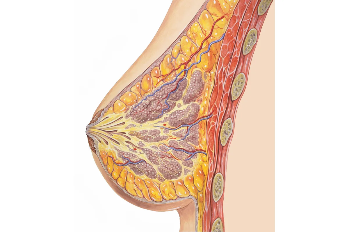
The breast lies on the front of the chest wall and is made of glandular tissue, milk ducts, fat and connective tissue, covered by skin. Breast tissue changes throughout life under the influence of hormones.
What is the structure of the breast?
Each breast contains about 15-20 lobes (gland compartments). Each lobe is made of many small milk-producing glands (lobules). Lobules are linked to the nipple by milk ducts, which join and open toward the nipple.
Fatty tissue and supporting connective tissue lie between the glands. Bands of connective tissue called Cooper’s ligaments attach the breast to the skin and the chest wall.
The nipple and areola
The nipple and the darker area around it (the areola) are sensitive skin. Small Montgomery glands in the areola keep the area moist. The milk ducts end at the nipple.
How do blood and lymph flow in the breast?
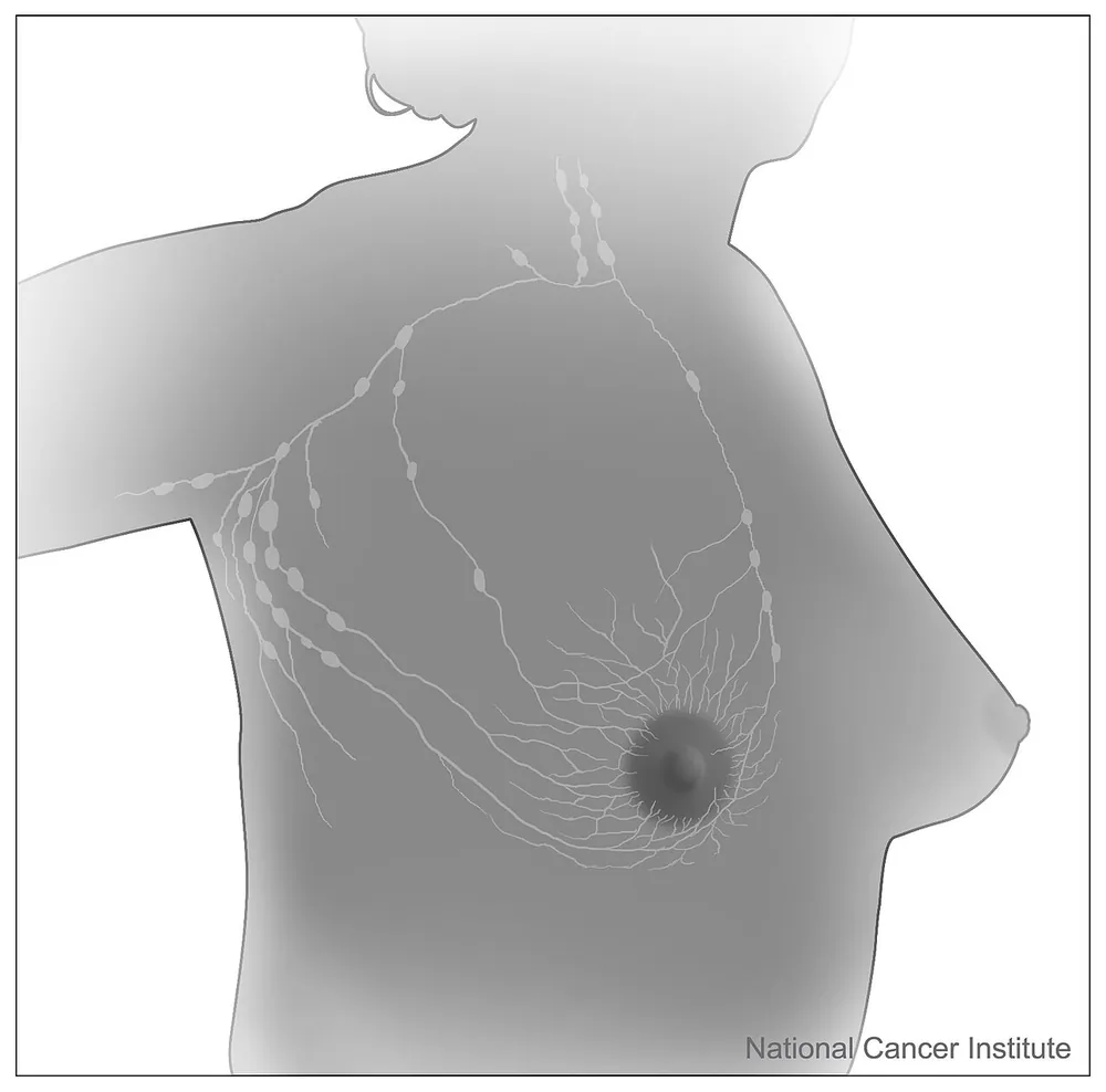
The breast is supplied by vessels of the chest wall and the armpit. Most of the lymph fluid drains to the lymph nodes in the armpit (axillary nodes), and some to nodes beside the breastbone and around the collarbone. This is why the armpit is also examined during a breast examination.
What lies beneath the breast?
Behind the breast are the pectoral muscle (pectoralis major) and the ribs. Breast tissue rests on top of the chest muscle.
Why is it important to know breast anatomy?
Knowing the normal structure of the breast makes it easier to notice changes during self-examination and forms the basis for imaging and surgical planning. If you notice a new lump, nipple discharge or a skin change, see a physician.
The content is for information purposes only and does not replace a medical examination.
Related pages: Breast Development, Breast self-examination, Physician examination
Images: Andrewmeyerson, CC BY-SA 3.0, Wikimedia Commons · Don Bliss / National Cancer Institute (public domain)